Radiology
Plan your appointment
What can we help you with?
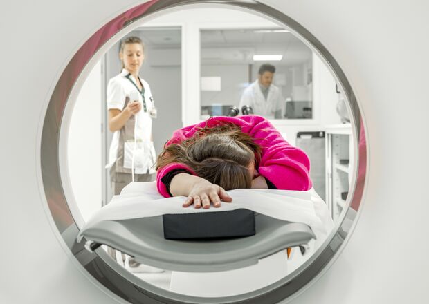
The service consists of several departments: classical radiology, mammography, ultrasound, CT scan, angiography with interventional radiology and NMR. Each of these techniques make their own contribution to imaging, and therefore several techniques are often complementary when examining a single organ. In many cases, a fluid is used that better distinguishes tissues from one another, called contrast fluids.
In the radiology department, both outpatients and hospitalised patients are examined.
Your radiology appointment
Be sure to always bring your doctor's request or prescription with you to your appointment at our radiology department.
Before you register at the radiology department, you must always enroll at the registration desk/registration kiosk in the entrance hall. For this, you need the following documents:
- your identity card
- the name of your GP and any home health care providers
- an insurance card or payment agreement from insurance (if applicable)
- a payment commitment from a CPAS (OCMW) or other agency that intervenes in the hospital bill (if applicable).
View your images online
The Nexuzhealth patient app makes it possible for you to access your medical records and view images of your examinations.
CLICK HERE to download the Nexuzhealth app or access your record through the website.
With the patient access code that you will receive at the radiology department, you can also view some images of your examination yourself.
To do this, click on the link below and enter your patient-access code and your date of birth:
Different types of research
RX examinations
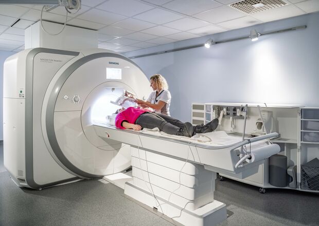
This type of examination is called conventional radiology or diagnostic radiology. In an RX examination, we take images of parts of the body based on X-rays. The result is called radiographs, on which abnormalities can be shown such as fractures, pneumonia, osteoarthritis, heart size, etc.
CT examinations
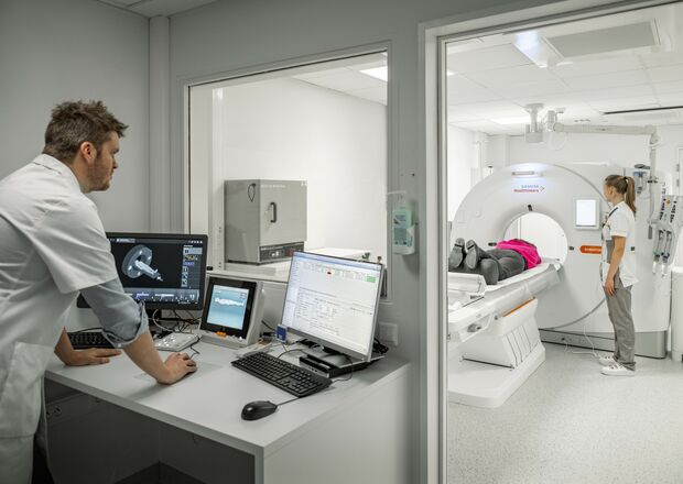
A CT-scan (computer tomography) forms an image of the body in a particular area by means of X-rays. Through this examination, we have the ability to detect abnormalities in certain organs.
> CT blood vessels (angio/cardio)
> CT chest (thorax)
> CT abdominal and pelvic organs (abdomen)
> CT limb
> CT skull
> CT virtual colonoscopy
NMR examinations (MRI)
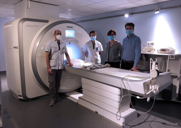
With an NMR (nuclear magnetic resonance), or MR(I), we image parts of the body by means of a magnetic field and radio waves. This is how we obtain 3D-images, disc images in three dimensions. No X-rays are used in an NMR examination. The examination can be considered harmless.
> NMR abdomen (abdomen)
> NMR blood vessels (angio)
> NMR bile ducts/pancreas (MRCP)
> NMR limb
> NMR mammo (breasts)
> NMR skull/brain
> NMR vertebral column
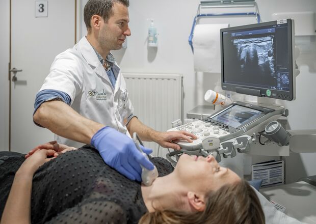
Echography
An ultrasound is an examination of certain body parts or organs that uses sound waves to look inside the body.
> Ultrasound breasts
> Ultrasound upper abdomen
> Ultrasound duplex
> Ultrasound limbs
> Ultrasound lower abdomen
Specific ultrasound examinations:
> Ultrasound with biopsy
> Ultrasound with spotting (harpoon placement)

Contrast studies
These are examinations in which we use contrast medium to get a good image of certain organs.
> Arthrography (joints)
> Colpocystodefaecography (pelvic floor for urinary and bowel movement problems)
> Contrast ultrasound
> Cystography (child)
> RX colon (large intestine)
> RX kidneys and urinary tract (IVP)
> RX esophagus
> RX esophagus, stomach and duodenum
> RX transit (small intestine)
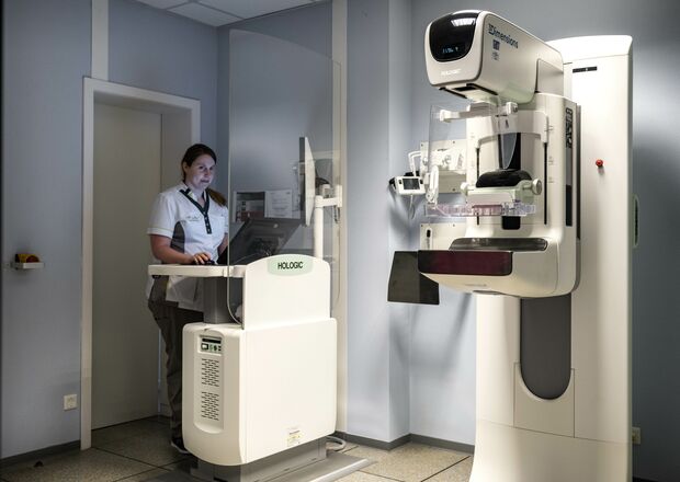
Breast examinations
Below you will find an overview of the different ultrasound examinations. Click on an examination for more detailed information.
> Ultrasound breasts
> Ultrasound with biopsy
> Ultrasound with spotting (harpoon placement)
> mammography
> mammotome biopsy
> NMR mammography
Other specific examinations
CT scan Cone Beam
Using a Cone Beam, our radiology department can make maxillofacial (face and neck) and dental imaging. This allows us to take three-dimensional, high-resolution images of the facial skeleton and tooth elements and do so with a dramatically lower radiation dose compared to the conventional CT scan.
Below is an overview of the different examinations with this device:
> CT scan Cone Beam
> Orthopantogram (OPG)
> Cephalogramme
Bone densitometry (DEXA)
A bone densitometry is a measurement of bone mineral density (BMD) at various places in the body such as the hip and spine and possibly the wrist or other bones. This examination uses a beam of very low-intensity X-rays to take images of these bones (hip, the lumbar spine column, the wrist) to measure the density of the bone and determine the degree of osteoporosis.
The examination can be performed using a DEXA device. This examination is completely painless and the amount of radiation is harmless. The examination takes only a few minutes.
> Bone densitometry DEXA
Treatments
Doctors
Head nurse
Application forms
Questionnaires
Where can you find us?
The radiology department is located in block A, follow the blue color from the entrance hall, all the way to the back of block A.
Before going to the radiology department, you must register at the registration kiosk in the entrance hall of the hospital.


